Introduction
The phrase “King Tut mummy face” captures both curiosity and scientific challenge. This article walks through what Tutankhamun’s preserved remains reveal about his facial anatomy, the techniques experts use to examine and reconstruct his look, and the limits of our knowledge. I write as an Egyptologist and forensic analyst to explain the evidence in clear, human terms.
Why the King Tut mummy face matters
Understanding the “King Tut mummy face” connects a real individual to ancient history. It helps scholars study health, genetics, mortuary practice, and how official royal images compare with biological reality.
Discovery and early examinations
Howard Carter found King Tut’s intact tomb (KV62) in 1922. The unwrapping and first examinations in the 1920s documented the mummy’s condition and began the long scientific conversation about the “King Tut mummy face.”
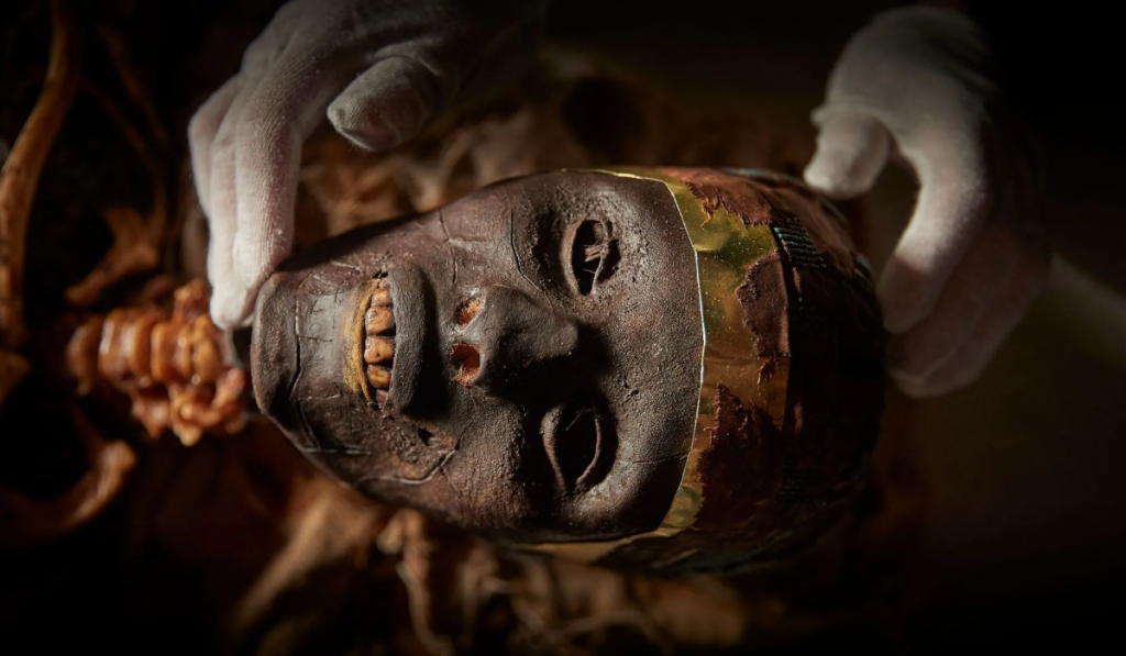
Condition of the mummy and its impact
Tutankhamun’s mummy shows desiccation, handling damage, and embalming alterations. These postmortem factors affect facial contours and must be considered when describing the “King Tut mummy face.”
Visible external facial features
- Narrower-than-expected facial proportions for a pharaoh.
- Slight midface asymmetry, visible on the preserved skull and in some reconstructions.
- Flattened soft tissues, typical of desiccated adolescent remains.
- Nose area altered by embalming materials and later damage.
- Dental pattern consistent with a late-teen age-at-death.
Skull and skeletal facial features
The craniofacial skeleton is gracile. Skull measurements show moderate midfacial projection and a nasal aperture consistent with ancient Egyptian populations. Small fractures from tomb handling exist, but no catastrophic skull trauma explains any dramatic facial deformity.
Distinctive facial characteristics
- Mild asymmetry: small left-right differences in cheek and jaw contour.
- Slight lower-jaw prognathism not strongly pronounced.
- Subtle healed lesions on the skull margin, likely postmortem.
Together these traits suggest a youthful, slender face rather than a heavily built or obviously deformed visage.
How age affects the King Tut mummy face
Tutankhamun likely died at about 18–19 years. Adolescent skull and soft-tissue proportions produce a jawline and cheek fullness different from a mature adult’s. Reconstructions therefore aim for youthful anatomy.
Methods used to study the “King Tut mummy face”
- Visual inspection during early unwrapping.
- Standard radiography to view bone beneath wrappings.
- High-resolution CT scans, which provide 3D models of bone, residual tissue, and packing.
- Endoscopy for minimally invasive internal inspection.
- Genetic and isotopic testing to contextualize ancestry and health (limits apply).
Combining these methods provides the best basis for facial description and reconstruction.
Forensic facial reconstruction techniques
- CT-based 3D skull modeling to form the skeletal foundation.
- Tissue-depth markers applied using population and age-specific datasets.
- Layered soft-tissue modeling: muscles, fat, and dermis built upon the skull.
- Nose, lips, and ears approximated from bony landmarks and comparative anatomy.
- Final rendering adds skin tone, hair, and expression inferred from genetic and cultural context.
Each step has uncertainty; reconstructions are informed approximations, not exact portraits.
Notable reconstructions of King Tut’s face
Early 20th-century wax and painted reconstructions reflected artistic norms. Modern digital reconstructions, derived from CT data, aim for anatomical precision. Museum displays vary in aesthetic choices—skin tone, hair style, and expression—so multiple plausible faces exist.
What CT scans specifically revealed
CT imaging clarified skull shape and dental status and separated embalming packing from biological tissue. Scans allowed precise cranial measurements and identification of packing that can mislead visual readings of cheek fullness.
Soft-tissue preservation vs. embalmer interventions
Actual soft tissue is sparse. Embalmers used resins, linen padding, and supports to shape the face for burial. CT scans are crucial for distinguishing embalming additions from the original soft tissues when reconstructing the “King Tut mummy face.”
Comparing iconography with anatomical reconstructions
Royal art idealizes rulers with symmetrical, ideal proportions. Tutankhamun’s golden mask projects ideal youth. Anatomical reconstructions show some overlap—youthful features—but also differences due to stylization and artistic conventions.
What the face suggests about health and heredity
Facial and skeletal evidence suggests a young person of slight build with possible inherited conditions in the royal lineage. Combined with limb abnormalities and other findings, the “King Tut mummy face” contributes to a picture of a royal with health issues, though not gross facial deformity.
Limitations, uncertainties, and interpretive choices
- Tissue depth standards are generalized and may not perfectly match ancient Egyptian adolescents.
- Genetic markers provide limited facial detail.
- Embalming and damage obscure original contours.
- Reconstructions require interpretive decisions; multiple versions are scientifically responsible.
Acknowledging limits is essential when presenting any “King Tut mummy face” reconstruction.
Ethical considerations and public presentation
Displaying reconstructions raises cultural and ethical questions about the treatment of human remains. Best practice requires transparency about methods, clear labeling of uncertainty, and sensitivity to descendant communities and cultural heritage.
Further reading and authoritative links
- Culture mosaic — general portal on Egyptian culture: https://culturemosaic.co.uk/
- The Golden Mummies of Egypt — feature article: https://culturemosaic.co.uk/the-golden-mummies-of-egypt/
- Most Famous Egyptian Mummies — comparative cases: https://culturemosaic.co.uk/most-famous-egyptian-mummies/
- Egyptian Concept of Life After Death — funerary context: https://culturemosaic.co.uk/egyptian-concept-of-life-after-death/
- Egyptian Mummy Iliad Papyri — textual context: https://culturemosaic.co.uk/5-egyptian-mummy-iliad-papyri/
- Where is king tut mummy — location and display history: https://culturemosaic.co.uk/where-is-king-tut-mummy/
- King Tut mummy face (Wikipedia overview): https://en.wikipedia.org/wiki/Tutankhamun%27s_mummy
Frequently Asked Questions About King Tut’s Mummy Face
FAQ 1 — How accurate is a reconstruction of the King Tut mummy face?
Answer: Reconstructions are well-informed approximations based on skull anatomy and CT data, but they are not exact likenesses.
- Use high-resolution CT as the foundation.
- Apply age- and population-appropriate tissue-depth standards.
- Record and publish all assumptions and methods.
- Compare outputs from multiple reconstruction techniques.
- Present reconstructions as interpretive, not definitive.
FAQ 2 — Can DNA reveal exactly how King Tut looked?
Answer: DNA can suggest ancestry and some pigmentation tendencies, but it cannot yet reconstruct a full facial appearance reliably.
- Use genetic data to inform pigmentation and ancestry cautiously.
- Combine DNA with cranial morphology for better estimates.
- Avoid categorical statements from single markers.
- Revisit reconstructions as genomic methods improve.
- Adhere to ethical rules for sampling ancient remains.
FAQ 3 — Did embalmers change the King Tut mummy face?
Answer: Yes. Embalmers used packing, resins, and linen that modified facial contours and must be accounted for.
- Use CT imaging to identify artificial packing.
- Discount heavily packed areas when modeling soft tissue.
- Consult funerary texts and comparative embalming cases.
- Run alternative reconstructions showing minimal and maximal packing effects.
- Note embalming influence in public-facing labels.
FAQ 4 — Which tools produce the most reliable facial reconstructions?
Answer: A combined workflow of CT-derived 3D skull models, validated tissue-depth datasets, and digital sculpting yields the most reproducible outcomes.
- Begin with volumetric CT segmentation of bone and residual tissue.
- Use validated tissue-depth markers for adolescents and regional populations.
- Employ layered digital modeling for muscle and fat.
- Validate with anthropometric measurements and peer review.
- Archive digital models and metadata for re-analysis.
FAQ 5 — How should museums present reconstructions of the King Tut mummy face?
Answer: Museums should present reconstructions as scientific interpretations, explain methods, show alternatives, and respect cultural concerns.
- Provide plain-language labels explaining methods and uncertainties.
- Display multiple reconstructions or an uncertainty range.
- Include contextual information about embalming and funerary beliefs.
- Involve ethical oversight and consult relevant communities.
- Provide links to primary research and further reading.